Control of stem cell divisions in the C. elegans epidermis
In the Caenorhabditis elegans epidermis, two lateral rows of “seam” cells form a polarized epithelium with stem-cell like divisions. These seam cells go through symmetric and asymmetric divisions with a reproducible anterior-posterior orientation at stereotypical times of development. We study these seam cells as an attractive model in which to uncover controls that determine the timing and plane of cell division, and the choice between symmetric or asymmetric division.
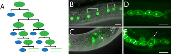
Epithelial seam cells as a model for polarity and asymmetric cell division. (A) Seam cell division pattern. Symmetric divisions result in a doubling of the seam cell number, while asymmetric divisions create a single seam cell and a cell that fuses with the epidermis. All divisions occur anterior-posterior. (B,D) Examples of markers used to follow seam-cell divisions. (B) Seam specific nuclear GFP. (D) Pleckstrin homology domain::GFP (cell membrane) and histone::GFP (DNA). (C,E) Disruption of polarity affects the number and localization of the seam nuclei. Scale bars are 10 µm.
We have created a number of transgenic reporter strains in which key cell division processes are visualized with fluorescently tagged proteins. We use these reporters in combination with time-lapse fluorescence microscopy, which allows following the position of the centrosomes and spindle, chromosomes, cell junctions, and cell membranes in the seam cells during larval divisions. Combined with forward and reverse genetics, this offers a powerful strategy for identifying regulators of the cell cleavage plane in an epithelial stem cell lineage. As many of the mechanisms are conserved across animal species, we expect that our findings in C. elegans will help understand the control of asymmetric cell division in human tissues (Wildwater et al. Development 2011).
Back to Overview