Welcome to the Braccioli Lab
Our goal is to identify novel gene regulatory mechanisms of tissue-tissue interaction using embryo models
We aim to dissect the physical and biochemical interaction between different organ primordia during mammalian embryonic development. We use stem cell-based embryo models (such as gastruloids) together with (single-cell) genomics, advanced microscopy, and reporter assays to study how gene regulation drives inter-tissue communication. Understanding the cross-talk between developing organs is a milestone towards the generation of organs in vitro for regenerative medicine.
Modeling early organogenesis in vitro
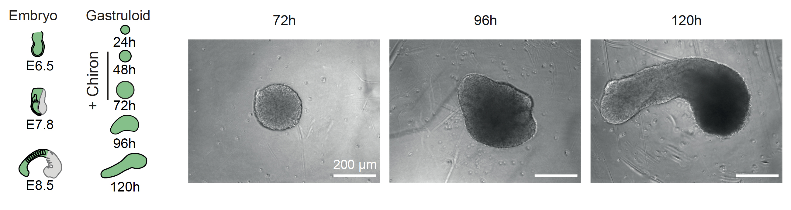
Gastrulation is a critical phase in embryonic development, characterized by the reorganization and differentiation of pluripotent cells into the three germ layers: endoderm, mesoderm, and ectoderm. This process establishes the body axes and initiates the formation of tissues that will develop into organs. These tissues are intricately interconnected and engage in continuous communication through physical contacts and biochemical signals, essential for driving cell fate decisions and morphogenetic processes. However, our understanding of the regulatory mechanisms driving inter-tissue communication during mammalian embryonic development is limited. This is due to the inherent challenges of manipulating and tracking embryos in utero. The recent advance of gastruloids, stem-cell based embryo models derived from pluripotent stem cells in vitro, allows to overcome these issues. Gastruloids are derived from aggregating a few hundred mouse embryonic stem cells (mESCs) and inducing their differentiation by treating them with the Wnt agonist Chiron. As they grow, they model many aspects of early trunk development, including germ layer specification, symmetry breaking and anterior-posterior axis formation. Importantly, they develop primordial tissues such as spinal cord, mesoderm, somites, cardiac progenitors, blood and vascular tissues. Gastruloids are highly scalable, easy to track, genetically engineer and manipulate. Moreover, they present a tractable tissue complexity when compared to more complex embryo models.
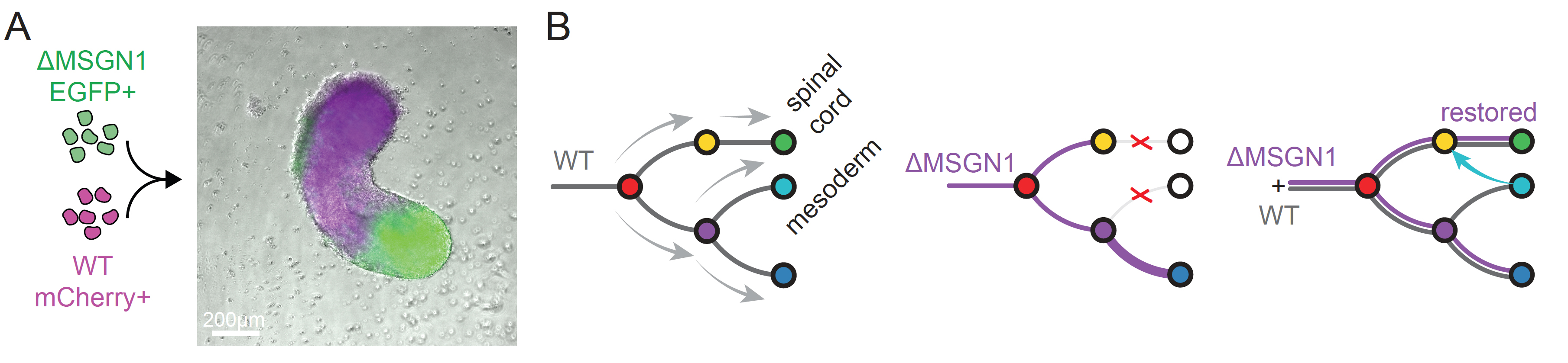
Dissecting inter-tissue communication with chimeric gastruloids
We recently developed the chimeric gastruloid model (panel A below), which is derived by aggregating gastruloids from a mix of wild-type (WT) and knockout (KO) cells labeled with different fluorophores (Braccioli et al. 2025). This allowed us to identify factors regulating inter-tissue communication. Therefore, we propose the chimeric gastruloid model as a powerful system to identify TFs driving inter-tissue communication and uncover the underlying gene regulatory programs. We previously delineated the tissue developmental trajectories and the TFs driving cell fate decisions in gastruloids by investigating the chromatin accessibility landscape in individual cells by single-cell (sc) Assay for Transposase-Accessibly Chromatin with sequencing (ATACseq) (Braccioli et al. 2025). Through this we predicted the TF MSGN1 to be active only in mesoderm cells. Surprisingly, we discovered that MSGN1 KO (ΔMSGN1) in gastruloids disrupted not only mesodermal cell development but also the development of the neighboring spinal cord cells. Using chimeric gastruloids we discovered that the presence of WT cells rescues the capacity of ΔMSGN1 to form spinal cord, highlighting an underlying gene regulatory program of inter-tissue communication driven by MSGN1 (panel B below). Using this innovative framework, we aim to discover novel mechanisms of inter-tissue communication.