Members
Gallery
Contact
Directions
Bachelor thesis
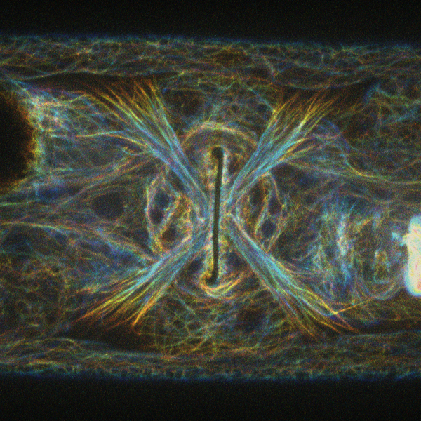
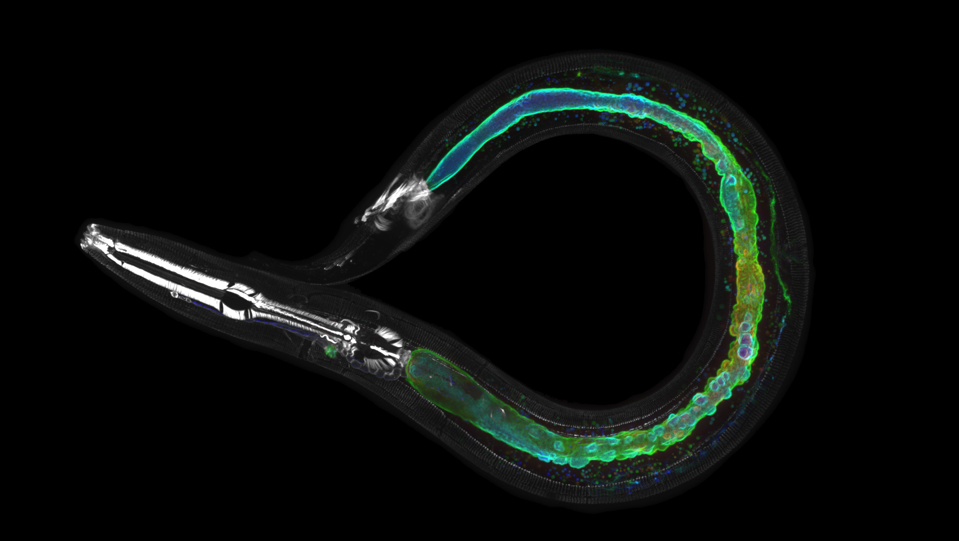
Ventral view of an adult animal. Endogenous GFP::MAPH-1.1 marks microtubules. Image false-colored by z-stack depth.Image by S. Waaijers
Endogenous GFP::BBLN-1 marks intermediate filaments(Image by Sanne Remmelzwaal)
Microinjection of a wild-type animal with a blue dyeInjection by H.R. Pires
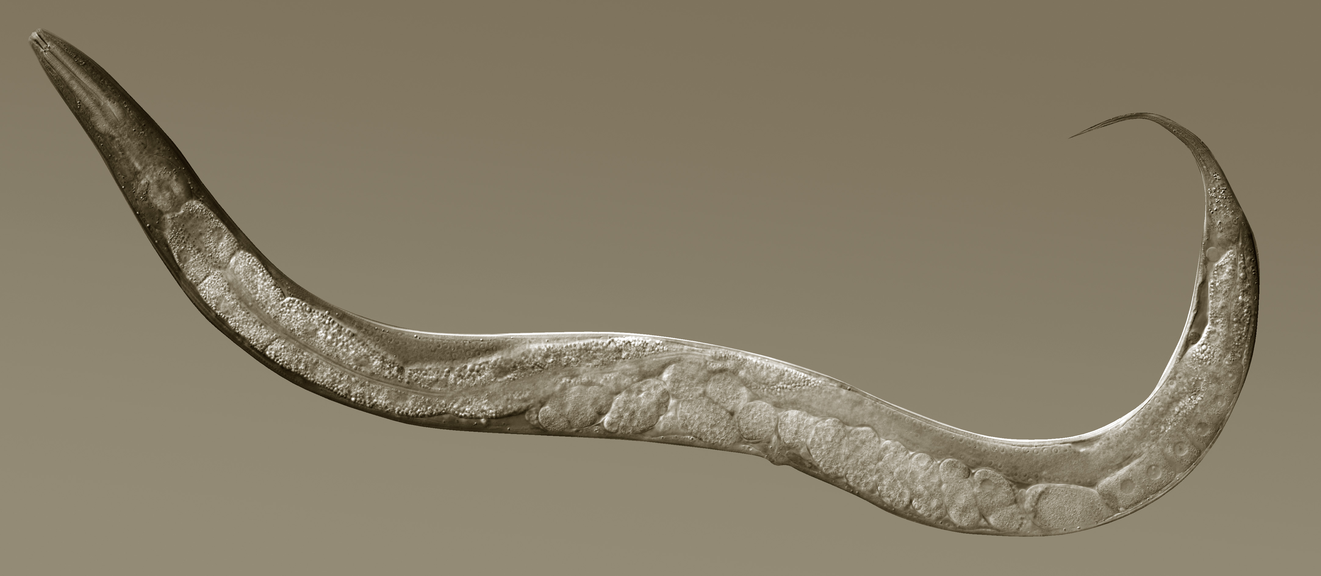
Nomarski DIC image of a wild-type N2 animalImage by M. Boxem
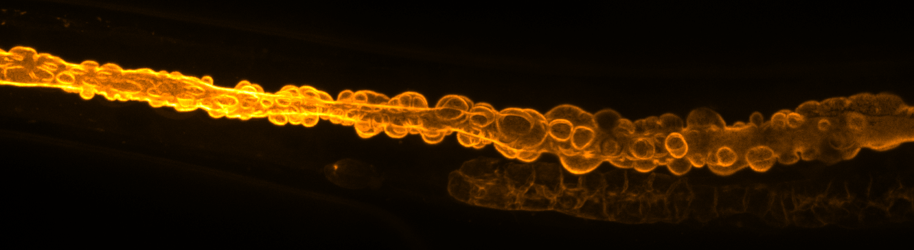
Apical membrane of the intestine labelled by ERM-1::GFP upon RNAi knockdown of bbln-1Image by J. Ramalho and S. Remmelzwaal
Expression of fluorescent markers in body wall muscle cells (purple) and intestinal nuclei (green)Image by M. Boxem
Worms being passaged – rather shakily – by M. Boxem
L4 larva expressing fluorescently tagged ERM-1 (colour coded by z-height) and the intermediate filament IFB-1 (gray) in a bbln-1 knockoutImage by Sanne Remmelzwaal
sqt-1 Roller worms expressing nuclear GFP from the sur-5 promoterVideo by L. Fielmich